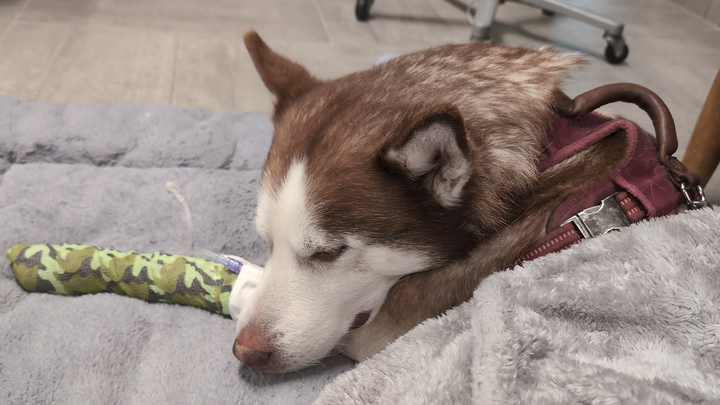
getting tests and medication for Levi
This is Levi and he is an 8 year old Husky. Suddenly his health started declining over the past week and he is in need of several tests to be able to figure out what is going on and hopefully to be able to save his life. Unfortunately, like many other Americans, we are extremely struggling financially and I have maxed out several credit cards already to be able to get the answers that we desperately need. Anything at this point will help us. This isn't supposed to be the end for him. I will include the results and estimated values from two different clinics for anyone who would like to see.
Update: They're heavily leaning towards IMHA (Immune-Mediated Hemolytic Anemia) or Cancer. There's most tests they want to run but are starting with medication to see if he reacts to it for IMHA.
1. When imaging the gall bladder, the radiologist did see an emerging mucocele. The function of the gall bladder is to emulsify fats; unfortunately, if it does not empty appropriately, the gall bladder can get too full and even rupture or block continued emptying. A medication called ursodiol can be used to help with gall bladder emptying to hopefully mitigate this problem. This is a medication that I can prescribe and you can get from a human pharmacy. Ideally, we like to recheck the gall bladder ( via ultrasound) after being on this medication after a few months to determine if there is improvement. Another option would be to surgically remove the gall bladder to prevent future rupture. This is a procedure that I would have to refer you to a specialty hospital for.
2. When imaging the prostate, the radiologist saw a paraprostatic cyst. Luckily, Levi is asymptomatic for this condition, but it does have the potential to cause abdominal pain, issues with urination etc. Ideally, surgical removal and neuter would be preferred prior to any clinical symptoms develop
I also received Levi?s lab samples back from the lab. Unfortunately, his labwork does look to be progressively getting worse rather than better. His anemia has worsened. He has gone from 33% to 29% (for reference normal range is 38-56% for dogs in Colorado), his albumin has decreased further from 2.0 to 1.9 (normal range is 2.7-3.9) and he continues to have electrolyte imbalances. I am concerned about these changes. It makes any surgery a higher risk for him. There are a few additional diagnostics we could perform to rule other conditions that may be causing th
: 1. Consider testing for Addisons ( hypoadrenocorticism) disease ( especially in light of his smaller adrenal gland on his ultrasound). This can be ruled out with a resting cortisol test ( ~ $102). If the test shows a cortisol higher than 2.0 then Levi does not have this disease. However, if it comes back low then we would have to perform a full ACTH stimulation test to confirm this disease (~ $
81). 2. Consider additional liver function testing such as a bile acids test (~$279). This test helps us determine how well the liver is functioning rather than just the appearance of the liver. Another potential diagnostic could be a liver biopsy but this is a surgical procedure as
well. 3. Consider additional imaging such as chest X-rays (~$330) to ensure nothing else is going on inside the chest for these c
nges. Once an anemia is below 25%, then we do consider blood transfusions in those patients. As well, with an albumin lower than 2.0, there is concern for Levi to develop fluid in his chest or abdomen. Therefore, I do consider these changes sig
ficant. This information can be overwhelming so if you would like to discuss this in more detail, I will be in the office all da
tomorrow.
Sincere
Dr. Kant Ultrasono
raphic Diagnosis: 1. There is a forming gallbladder mucocele. Surgical removal of the gallbladder could be performed. Alternatively, consider the use of ursodiol in this patient. If this medication is used, warn the owners that in rare instances, this medication can result in gallbladder rupture and/or b
liary obstruction. 2. The appearance of the prostate is most likely due to benign prostatic hyperplasia since this is an intact male dog. Prostatitis or prostatic neoplasia are unlikely. There is a paraprostatic cyst originating from the left lobe of the prostate. Surgical removal of the paraprostatic cyst is recommended. If this patient is not used for breeding or in shows, neute
ing is recommended. 3. The hyperechoic foci in the testes is most likely a result of dystrophic mineralization and much less likely due to neoplastic mineralization. The heterogeneity of the testicular parenchyma could be a normal aging change, associated with decreased sperm production, or a result of carcinoma in situ. Again, neutering of this pa
ient is recommended. 4. The small amount of peritoneal effusion may be a transudate, modified transudate, hemorrhage, or
unlikely, an exudate. 5. The overall appearance of the liver may be due to infiltration of fat, fibrin, or glycogen (aging changes), infectious/inflammatory disease (chronic active hepatitis, cholangiohepatitis, or other non-specific hepatopathy, copper storage disease, toxin ingestion, or infiltrative neoplasia. Bile acids testing or fine needle aspiration or, preferably, biopsy of the liver may yield
definitive diagnosis. 6. The hyperechoic foci in the spleen are likely the result of dystrophic mineralization or siderotic plaques as no other sonographic abnormalities
are seen in the spleen. 7. The material in the urinary bladder may be fat, crystals, or other cellular debris. A urinalysis and/or urine culture may b
useful in this patient. 8. The subjectively small size of the left adrenal gland may be a normal variant for this patient. While unlikely, a resting cortisol could be done to rul
out hypoadrenocorticism. 9. A cause for the reported anemia, hypoalbuminemia, and high cholesterol is not found on this examination unless it is related to one of the above findings (e.g. liver disease as gastrointestinal and renal disease do not appear likely on this examination) or is hereditary. Quiz the owners if there are any gastrointestinal clinical signs that may support protein losing enteropathy despite the normal sonographic appearance of the gastrointestinal tract and/or consider a diet trial with a highly digestible, hypoallergenic, or hydrolyzed diet, a gastrointestinal panel sent to Texas A&M University, and/or full thickness biopsies of the gastrointestinal tract. Thoracic radiographs could be done to rule out pathology in the thorax, but otherwise consider immune-mediated disease, infectious/inflammatory disease, toxin ingestion, or bone marrow disease as
