Pupeh needs another surgery
Donation protected
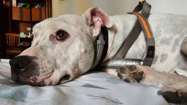
Hi, this is Pupeh. He came to our lives on April 2014 in Dublin, Ireland, he was a approximately 6 months. Apparently he was a Christmas present for someone and when they realised that was deaf we was completely neglected. Then some nice people appeared and fostered him ( Angie, Laura and Clondalkin Dogs Aid) even tracked his brothers, that were also deaf rescued and rehomed them.
He is the kindest dog, Lars' big brother and my best friend.
We moved to Latvia, and then here, Barcelona, he's been always by my side, more than moral support, the best companion and the sweetest of the souls.
If you know me, you know this year has taken a toll on me emotionally and economically, in April I had to emergency travel home (Argentina) due to a family emergency that wouldn't have been possible without the help of some amazing people, I was there for 55 days. And just after a week back here sadly, my dad passed.
Upon my return, I found a pimple, that didn't look good on Pupeh's belly, he was treated with 3 different things for 3 weeks, then reached the decision to have it removed and have it biopsied. Unfortunately the biopsy didn't come back the way we expected, and the one option he has is to have another surgery.
(BIOPSY details below)
The plan now is to have a surgery to remove the lymph nodes before gets too aggressive and start causing trouble, another biopsy after that, antibiotics, prebiotics, and keep him on the immune support meds.
I just want to help him stay here longer, without suffering.
If you can help or share we'd be beyond thankful.
BIOPSY DETAILS
Species: Dog (Canis lupus) Breed: American Staffordshire Terrier Sex: Castrated Age: 9 year/s, 0 month/s Sample type: Biopsy Correct Registration date: 07-22-2023 Validator: Francisco Fernandez Flores DVM, PhD (07-31-2023) SIMPLE BIOPSY (No. of Tissues: 1) Francisco Fernandez Flores FABRIC 1 Sample type: Skin lesions located between the base of the penis and the area near the nipple Macroscopic description: Cutaneous tissue of 4.3 x 2.5 cm with 1 nipple and nodules of 1.2 cm Ø (1) and of 0.6 cm Ø (2) Microscopic description: (1) (2) - The sections obtained from both lesions show the development of a proliferation multilocular neoplastic, infiltrative, derived from the epidermis, composed of epithelial-type cells organized in partially stratified nests, of variable size, with central scaly differentiation and centers occupied by keratin laminar (corneal pearls). The cells have a polygonal morphology, broad cytoplasm, broad, eosinophilic or hypereosinophilic, and a large, severely anisokaryotic, rounded or oval nucleus, with reticulated or granular chromatin and a or several prominent nucleoli. The mitotic count is moderate (1-3 mitoses/40x field), with the presence of mitoses atypical Dorsally, in the largest lesion the epidermis is severely ulcerated. In the rest of the tissue, so diffuse, the epidermis shows moderate irregular hyperplasia with polypoid elevations and digitation formation multifocal from which small neoplastic nests arise with characteristics similar to those described previously The changes are accompanied by hyperpigmentation of the stratum basale and pigmentary incontinence with melanomacrophages and moderate mixed inflammatory infiltrate, predominantly lymphoplasmacytic, with associated neutrophils in areas of ulceration, edema and subepithelial hemorrhages. The lymphatic vascular component associated with the evidenced lesions, shows the multifocal presence of emboli intravascular neoplastic cells. Mitotic count: 21 in 10 fields hpf / 2.37 mm² Lymphovascular invasion: Presence Necrosis: 15-20% of the total analyzed Surgical margins Lateral margins: Absence of cells in contact with the margins (1-2 mm at the surgical margin) Deep margins: Absence of cells in contact with the margins (2-3 mm at the surgical margin) Diagnosis: (1) (2) - SQUAMOUS CELL CARCINOMA, HIGHLY INFILTRATING, WITH INTRA AND PERILERSIONAL LYMPHATIC CARCINOMATOSIS.
Diagnosis: (1) (2) - SQUAMOUS CELL CARCINOMA, HIGHLY INFILTRATING, WITH INTRA AND PERILERSIONAL LYMPHATIC CARCINOMATOSIS. General observations: The lesional characteristics are indicative of the development of a malignant neoplastic process originating from of the stratified lining epithelium, with locally infiltrative behavior. Although the injuries are within of the specimen resection margins, the recurrence rate of these lesions after surgical excision is high, taking into account the multifocal nature of the proliferation together with the presence of multifocal preneoplastic changes at the epidermal level. On the other hand, the location of the lesions identified in this case (peripreputial area) is relatively uncommon in dogs and its association with some etiological factors such as papillomavirus infection or sun exposure is uncertain. The images of lymphangiosis examined are indicative of implanted or imminent metastases. We recommend one strict monitoring of the patient in the short term, monitoring the condition of the lungs and regional lymph nodes closest to the injury as well as considering the possibility of performing a radical excision associated with regional lymphadenectomy, if lymphnodular involvement is identified. Likewise, it is recommended to avoid sun exposure as much as possible, since exposure to UV rays can favor the development of this type of neoplasms in poorly pigmented areas..
Organizer
Marian Doratto
Organizer
Barcelona, CT